Team : STUDER
Research Projects
3D multicolor imaging
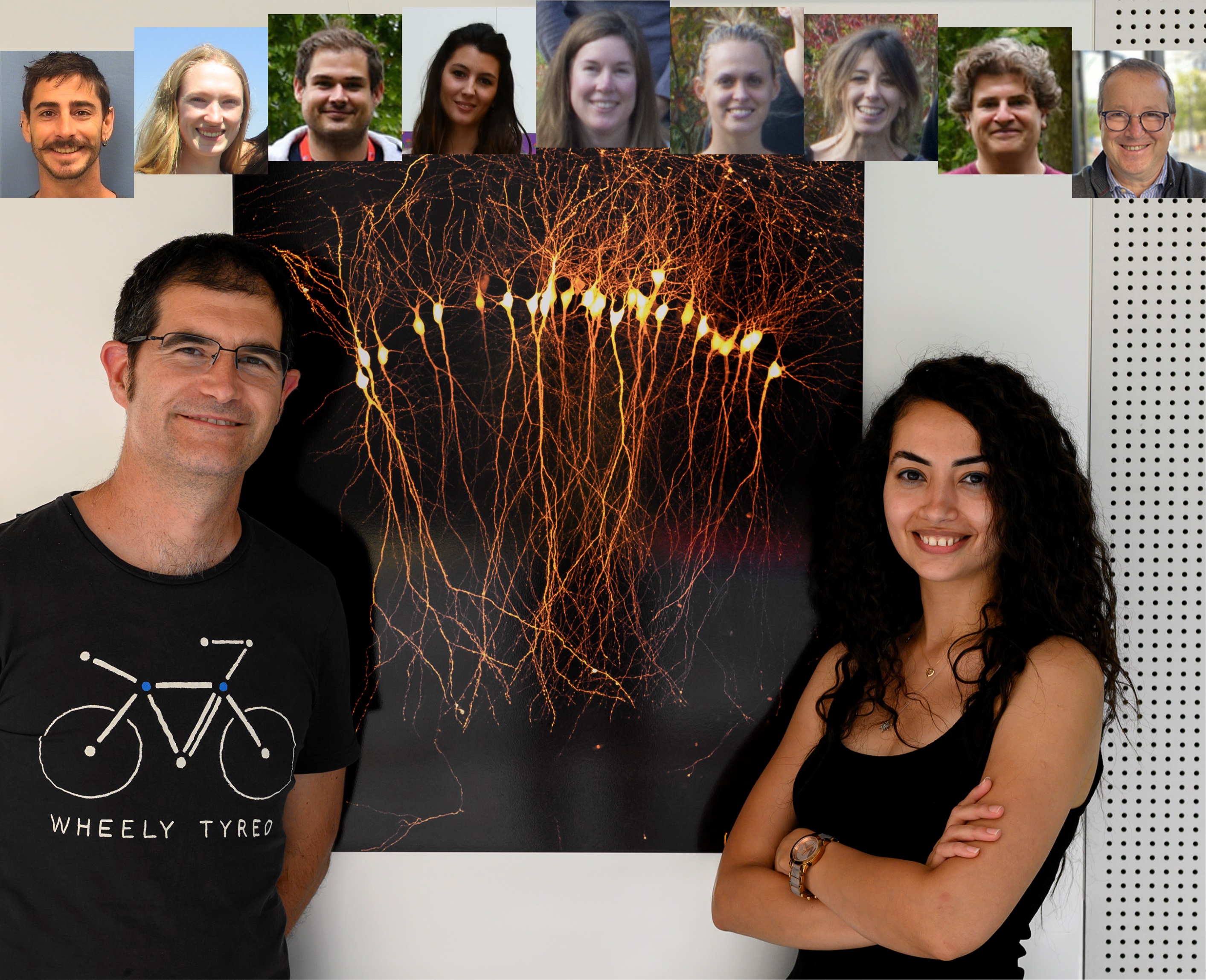
Project Leader(s): Vincent Studer / Sarah rahmati
One of the major challenges in imaging microscopy in 3D is to maintain viability of cells following prolonged exposure to excitation illumination to minimize photobleaching and photodamage over the course of the acquisition time.Our approach to perform long term 3D imaging is Image Scanning Microscope (ISM). ISM is a combination of Laser Scanning Confocal Microscope and an array detector (Camera). A multifocal illumination pattern is generated by a digital micro-mirror-device (DMD) that scans the sample over to provide a sectioned image of the focal plane in 1 second at the resolution of laser scanning confocal microscope.
To achieve simultaneous multiple color imaging the signal of different wavelength beam is separated onto the camera array using a prism. The wavelength of the beam is slightly deviated from the optical path at each illumination point of the grid, leading to multiple intensity peaks onto the camera.This technique enables long term imaging of brain tissue and organoids in 4D.
We have various projects based on the use of this imaging system including a project in collaboration with Matthieu Letellier (IINS- Thoumine Team) on the study of synaptic plasticity on organotypic slices with single electroporated neurons.
Fundings
None